Instrumentation and Image Processing
I’m Samuel Davis, a postdoc in the Caltech Optical Imaging Laboratory under the supervision of Dr Lihong V. Wang. I work on the development of photoacoustic imaging instrumentation and image processing. I did my PhD at Dr Paul French’s photonics lab at Imperial college London, developing optical projection tomography.
Photoacoustic Computed Tomography
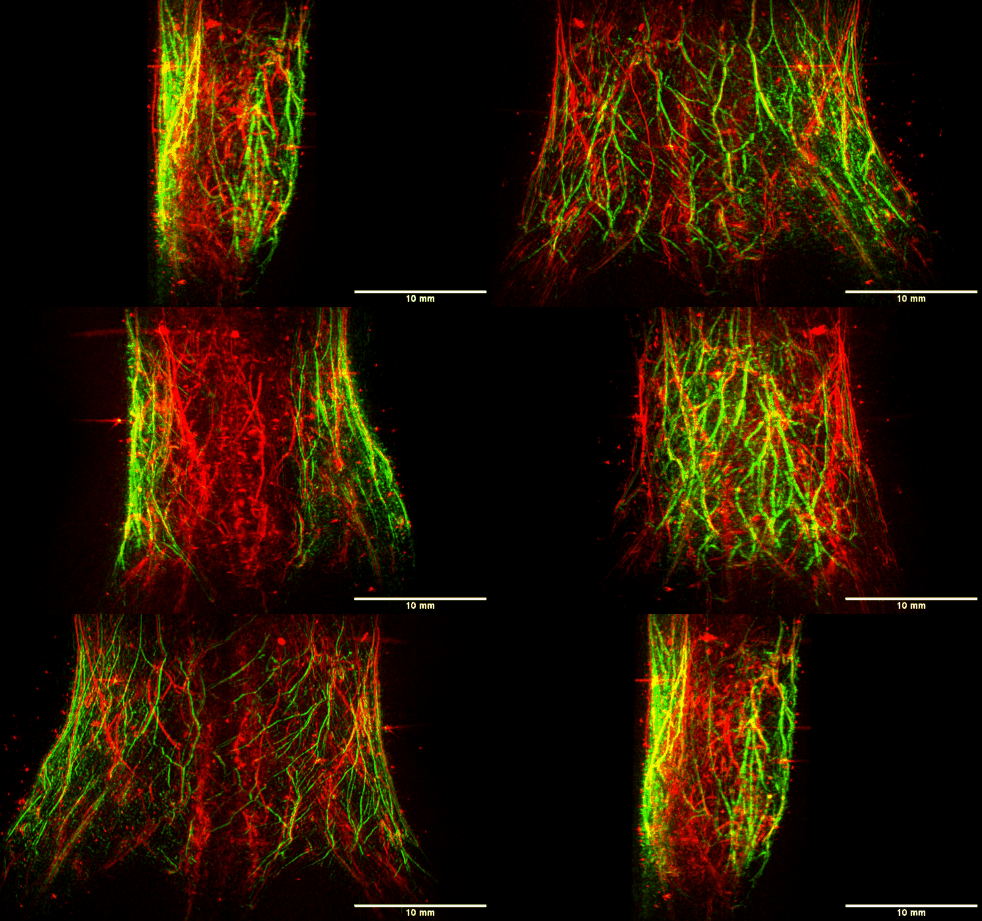
Photoacoustics is an emerging technology that enables optical contrast imaging beyond the diffusion limit by using acoustic detection. I work on the development of the next generation of pre clinical imaging instrumentation. Smaller resolution requires higher ultrasound frequencies, which suffer from stronger attenuation in tissue and water. Developing these systems is a challenge of maximising signal and minimising noise
Unsupervised Deep Denoising
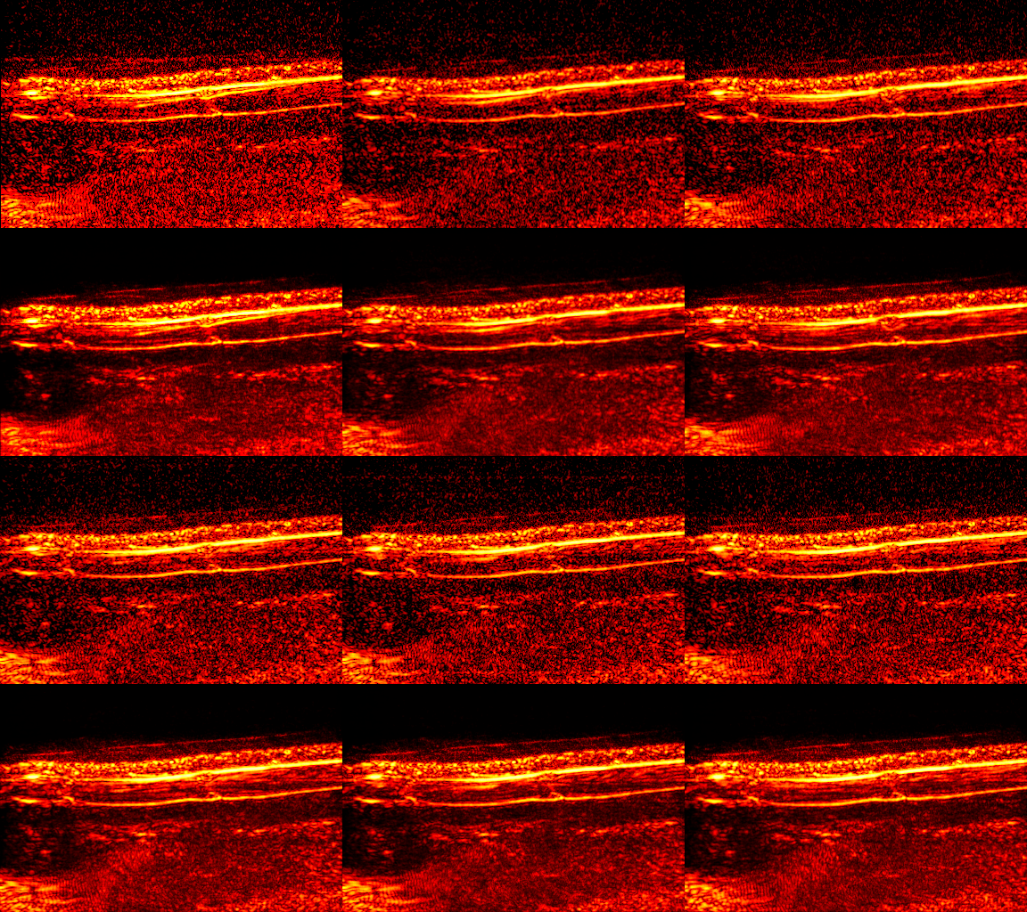
One of the main drawbacks of deep learning is the need for lots of high quality training data. A research interest of mine is finding self supervised methods that require no labels and/or can be trained using only the data of interest. Here, a network is trained to predict video frames from previous frames. It is able to learn correlated signals, but has too few parameters to learn high entropy noise - producing a video with reduced noise
Psuedo Colour for Photoacoustic Microscopy
Photoacoustic microscopy can be used to rapidly image challenging pathology samples, such as bone, that require up to a week of preparation for conventional imaging. This allows for intraoperative imaging of tumour margins - reducing the amount of healthy tissue that must be removed. To aid pathologist interpretation we developed self-supervised virtual staining using cycleGAN - an architecture that uses a self consistency loss to remove the need for well matched training data.
Optical Projection Tomography
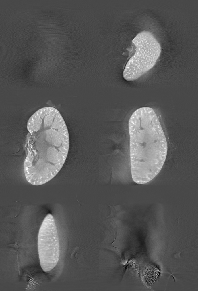
Optical projection tomography (OPT) is the optical analogue of Xray computed tomography. OPT systems are low cost - any widefield microscope can perform 3D imaging with the simple addition of a rotation stage. During my PhD I worked on the development of OPT instrumentation and image reconstruction, including a single pixel camera for low cost short wave infrared imaging. This is a reconstruction of the autofluorescence signal from a mouse kidney.
Undersampling artifact removal
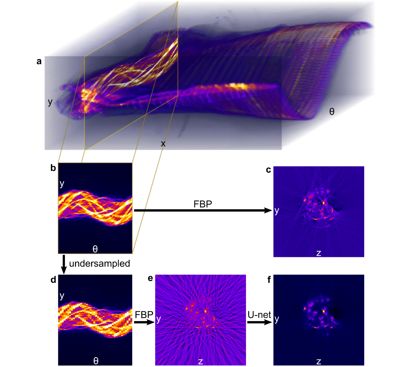
For in vivo imaging we want to minimise data acquisition times. However if we sample too few projections we get streak arifacts in our reconstructions. These can be used using compressive sensing using total variation regularisation, but we can get better results using machine learning. To demonstrate the robustness of the streak reduction we trained the network using data from ex vivo mouse tissue, and applied it here to in vivo zebrafish data.
